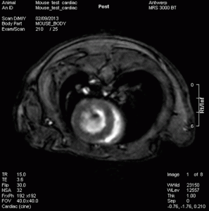
Cardiovascular

Cardiovascular magnetic resonance imaging (CMR), sometimes known as cardiac MRI, is a medical imaging technology for the non-invasive assessment of the function and structure of the cardiovascular system. It is derived from and based on the same basic principles as magnetic resonance imaging (MRI) but with optimization for use in the cardiovascular system. These optimizations are principally in the use of ECG gating and rapid imaging techniques or sequences. By combining a variety of such techniques into protocols, key functional and morphological features of the cardiovascular system can be assessed.
Cardiac MRI creates both still and moving pictures of the heart and major blood vessels. Doctors use cardiac MRI to get pictures of the beating heart and to look at its structure and function. These pictures can help them decide the best way to treat people who have heart problems.
Ideal technique for:
- Coronary heart disease
- Damage caused by a heart attack
- Heart failure
- Heart valve problems
- Congenital heart defects (heart defects present at birth)
- Pericarditis (a condition in which the membrane, or sac, around your heart is inflamed)
- Aneurisms
- Cardiac tumors
- Cardiomyopathy
References:
http://www.nhlbi.nih.gov/health/health-topics/topics/mri/
http://en.wikipedia.org/wiki/Cardiac_magnetic_resonance_imaging

