Image Gallery
-
Learn more

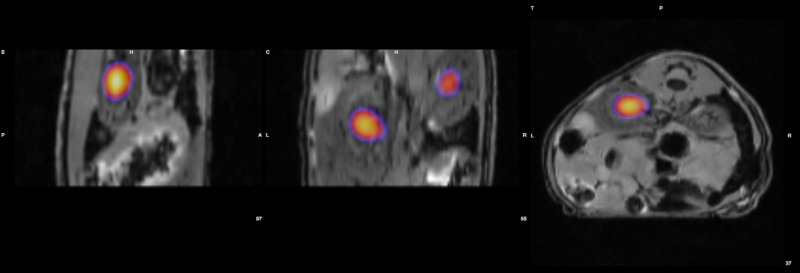
Mouse Multi-modality Imaging (Optical-MR)
Hectotopically injected colo-205 tumor cells
Kidneys MR + Bioluminescence
MR
FLASH 3DImage courtesy of: Prof Annemie Van der Linden, Bioimaging Laboratory, University of Antwerp
Learn more

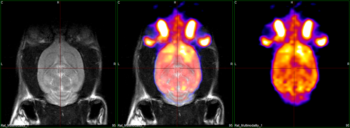
Rat Brain Multi-modality Imaging (PET-MR)
RAT
Strain: CD (SD) / Gender: female
Age: >6 months / Weight: 400gPET
Isotope: 18F / Dose: 31Mbq IV
Uptake: 45min
Acq. mode: Static
Energy Window: 250-700 KeVMR
FSET2WOrientation: Axial
Slice thickness: 1mm / 18 slices
TR: 3000ms / TE: 68ms
FOV: 35×35 / FrxPh: 256×248 / Averages: 4
Acq. Time: 6:16minImage courtesy of: Prof. Brunotte, FCS, Fondation de Coopération Scientifique du PRES Bourgogne Franche-Comté, Centre George-François Leclerc, Oncodesign CNRS Dijon. This work received support from the French Government managed by the French National Research Agency under the program “Investissements d’Avenir” with reference ANR-10-EQPX-05
Learn more

Mouse Brain Multi-modality Imaging (PET-MR)
Control mouse with middle cerebral artery (MCA) occlusion. Normal and high uptake patterns predict minor stroke, mixed and low patterns predict moderate stroke, and absent perfusion pattern is a predictor of severe stroke and death.
MOUSE
Strain: CD1 / Gender: female
Age: >6 months / Weight: 30g
Imaged 8h after MCA permanent occlusionPET
Isotope: 18F / Dose: 25Mbq IP
Uptake: 30min
Acq mode: Static
Energy Window: 250-700 KeVMR
FSET2WOrientation: Axial
Slice thickness: 1mm / 16 slices
TR: 4000ms / TE: 68ms
FOV: 30×30 / FrxPh: 256×248 / Averages: 4
Acq. Time: 8:21minImage courtesy of: Prof. Brunotte, FCS, Fondation de Coopération Scientifique du PRES Bourgogne Franche-Comté, Centre George-François Leclerc, Oncodesign
CNRS Dijon
This work received support from the French Government managed by the French National Research Agency under the program“Investissements d’Avenir” with reference ANR-10-EQPX-05
